OpenRoentgen
Web app
Coming soon, In development
A radiology
knowledge base.
Recorded OpenRoentgen demo: a question about Warthin tumor versus pleomorphic adenoma, followed by a cited answer and published MRI, CT, and ultrasound examples. The recording tours its images and references automatically.
Which imaging features help distinguish Warthin tumor from pleomorphic adenoma of the parotid?
- 2 images · 1 article passage · 2 abstracts · Source check passed
Ultrasound and MRI provide several distinguishing features, although the reported findings vary by modality and study. On gray-scale ultrasound, lobulated margins were noted in 84.2% of pleomorphic adenomas and 51.7% of Warthin tumors, while anechoic cystic components occurred in 44.8% of Warthin tumors and 5.3% of pleomorphic adenomas in one retrospective study.1
On MRI, pleomorphic adenomas in one study mostly showed T2-hyperintense solid components, whereas moderate T2 signal in solid components was more common in metaplastic Warthin tumors; cyst/necrosis was also more common in metaplastic Warthin tumors than pleomorphic adenomas.2 In that study, most pleomorphic adenomas showed marked enhancement, while 52.4% of metaplastic Warthin tumors showed moderate or marked enhancement.2
DWI and dynamic contrast-enhanced MRI can add useful distinctions: a 10-Warthin/13-pleomorphic adenoma study found lower ADC and higher tumor blood flow in Warthin tumors; many Warthin tumors had early enhancement with high washout, while many pleomorphic adenomas had gradual enhancement.3 In that cohort, reported diagnostic accuracies were 87.0% for ADC, 95.7% for tumor blood flow, and 91.3% for visual analysis.3
Parotid Pleomorphic Adenoma
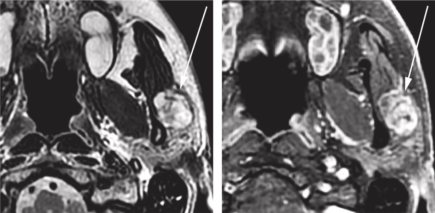
MRI · Baseline 3 T neck MRI without and with gadolinium
Source caption61‐y‐old man with benign mixed tumor (pleomorphic adenoma) of the left parotid gland. MRI of the neck with and without gadolinium‐based contrast reveals a well‐defined, T2‐hyperintense (A), heterogeneously enhancing (B) lesion in the left parotid gland. Minor T2‐hypointense septa are noted within the lesion
Library example · Ref. 4 · PMC7813040
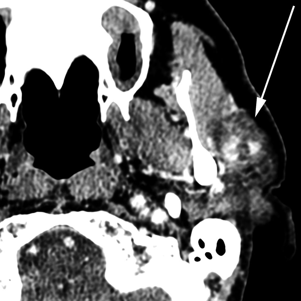
CT · Initial contrast-enhanced neck CT
Source caption61‐y‐old man with benign mixed tumor (pleomorphic adenoma) of the left parotid gland. Initial CT of the neck with contrast reveals a heterogeneously enhancing, relatively hypointense, well‐defined lesion in the left parotid gland measuring 2.6 × 2.1 × 1.9 cm
Library example · Ref. 4 · PMC7813040
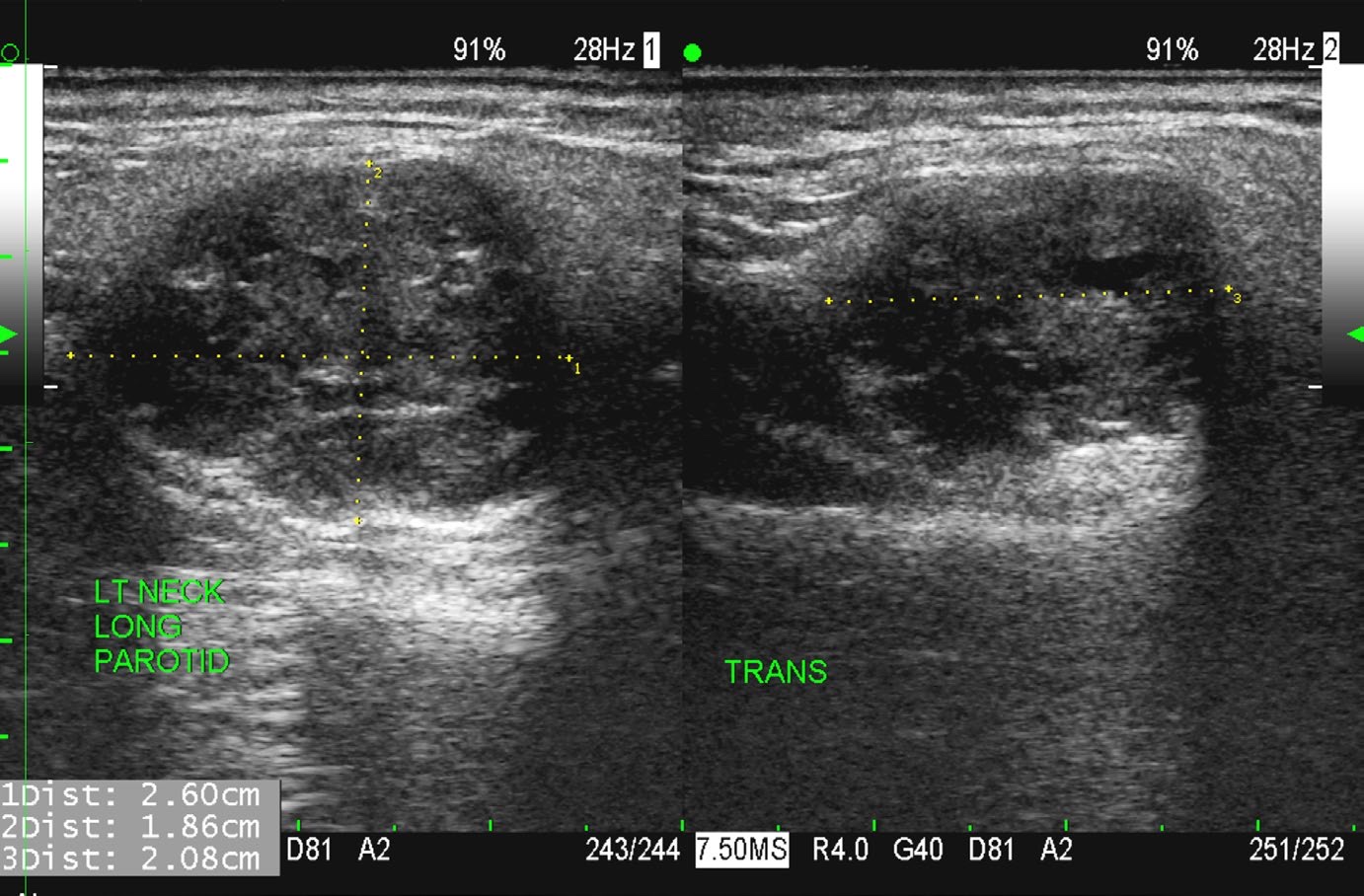
Ultrasound · Left parotid ultrasound
Source caption61‐y‐old man with benign mixed tumor (pleomorphic adenoma) of the left parotid gland. Ultrasound of the left parotid lesion reveals a slightly heterogeneous echotexture but a predominantly hypoechoic lesion with well‐defined borders and mild posterior acoustic enhancement. The lesion size measured with ultrasound, 2.6 × 2.1 × 1.9 cm, agreed with the size measured with CT
Library example · Ref. 4 · PMC7813040
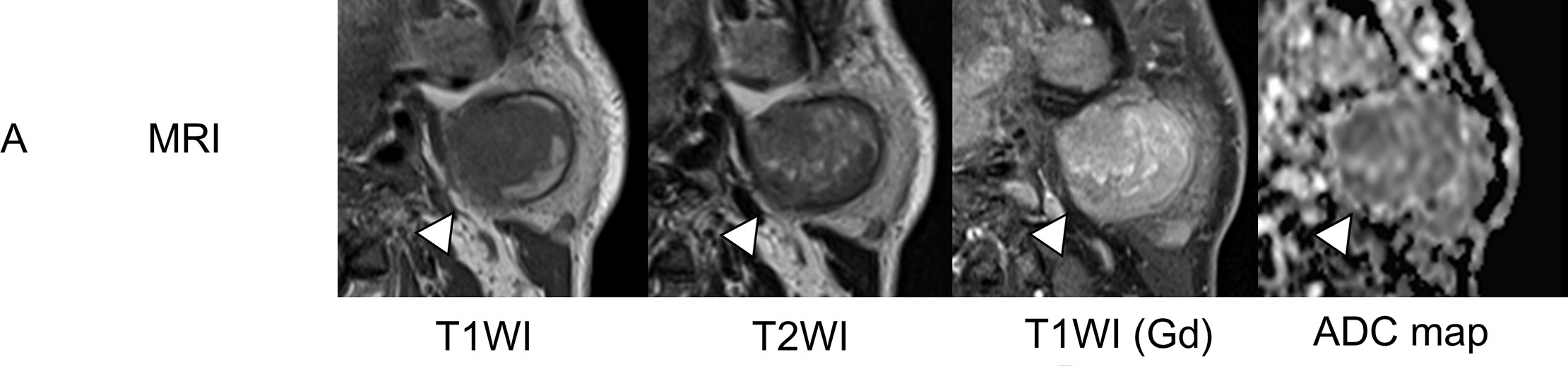
Warthin Tumor · Axial parotid MRI with gadolinium and ADC map
Source captionWarthin tumor on the left side of a 60-year-old man (case 14). (A) Axial MRI (white arrowheads) showed heterogenous (low to high) intensity on T1WI, heterogenous (low to intermediate) intensity on T2WI, strong gadolinium enhancement, and a low signal on the ADC map (ADCmean 0.94 × 10⁻³ mm2/sec).
Library example · Ref. 5 · PMC4744013
References
- 1.
Yuan, Wei-Hsin; Hsu, Hui-Chen; Chou, Yi-Hong; Hsueh, Huai-Cheng; Tseng, Tse-Kai; Tiu, Chui-Mei. Gray-scale and color Doppler ultrasonographic features of pleomorphic adenoma and Warthin's tumor in major salivary glands. Clinical imaging. 2009 PMID 19712813 · Journal Rounds abstract
- 2.
Jia CH, Wang SY, Li Q, Qiu JM, Kuai XP. Conventional, diffusion, and dynamic contrast-enhanced MRI findings for differentiating metaplastic Warthin’s tumor of the parotid gland. Science progress. 2021. doi:10.1177/00368504211018583 PMC10455002
- 3.
Yamamoto, Tatsuya; Kimura, Hirohiko; Hayashi, Kuniyoshi; Imamura, Yoshiaki; Mori, Masaki. Pseudo-continuous arterial spin labeling MR images in Warthin tumors and pleomorphic adenomas of the parotid gland: qualitative and quantitative analyses and their correlation with histopathologic and DWI and dynamic contrast enhanced MRI findings. Neuroradiology. 2018 PMID 29968073 · AbstractTop radiology journal
- 4.
Johnson JM, Mohamed ASR, Ding Y, et al. Ultra‐small superparamagnetic iron oxide (USPIO) magnetic resonance imaging in benign mixed tumor of the parotid gland. Clin Case Rep. 2020;9(1):123–7. doi:10.1002/ccr3.3477 PMC7813040 · CC BY 4.0 · Image source · License
- 5.
Kikuchi M, Koyasu S, Shinohara S, Imai Y, Hino M, Naito Y. Preoperative Diagnostic Strategy for Parotid Gland Tumors Using Diffusion-Weighted MRI and Technetium-99m Pertechnetate Scintigraphy: A Prospective Study. PLoS One. 2016;11(2):e0148973. doi:10.1371/journal.pone.0148973 PMC4744013 · CC BY 4.0 · Image source · License
Recorded demo · the composer is inactive here.
Images: Johnson et al., 2020 · Kikuchi et al., 2016 · CC BY 4.0.
Ask a radiology question and get an answer with citations and images from the medical literature.